Inferiorly it is reaching the hyoid bone with no erosion or related extensions.
Floor of mouth radiopaedia.
The pharyngeal fossa or foveola pharyngica is a variant oval or round depression within another inconstant bony excavation the fossa navicularis on the ventral surface of the basioccipital clivus.
Herein we have an oral cavity neoplasm that appears to originate from the anterior tongue to spread into the floor of mouth invading structures as described.
Knowledge of the anatomy of the oral cavity is imperative to understand the site of origin of the neoplasm the pattern of spread and enables documenting the stage of the disease.
Cysts superficial to geniohyoid may cause posterosuperior displacement of the tongue dysphonia dysphagia 3 or airway obstruction 4.
Angina ludovici is a type of severe cellulitis involving the floor of the mouth.
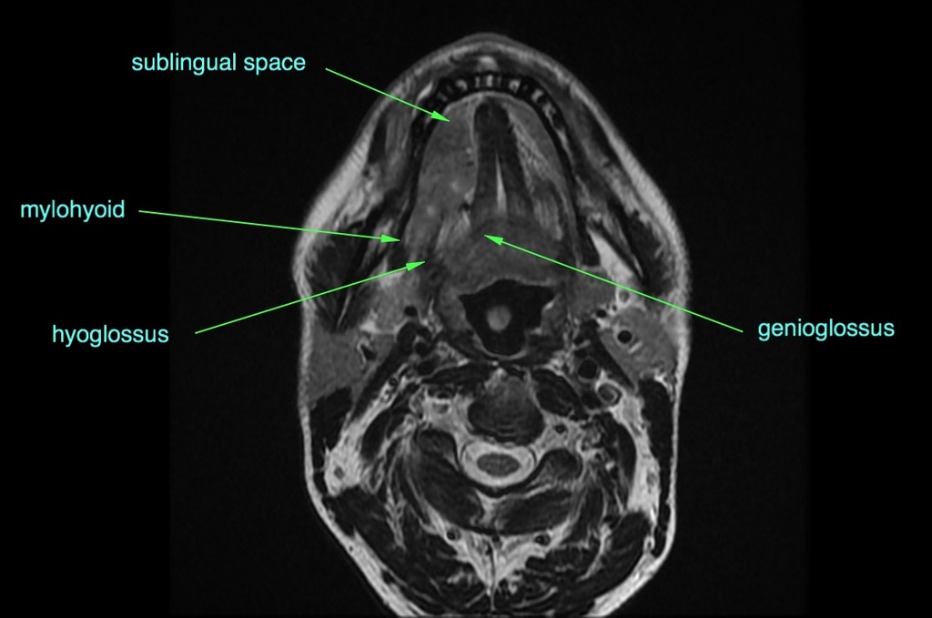
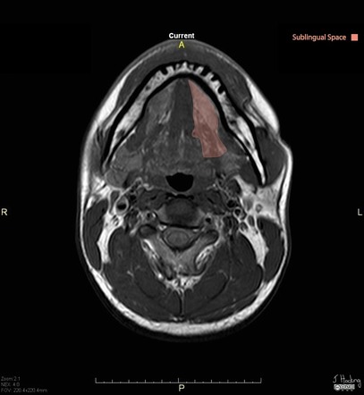
The floor of mouth is a u shaped space which extends and includes from the oral cavity mucosa superiorly and the mylohyoid muscle sling 2 3.
Early on the floor of the mouth is raised and there is difficulty swallowing saliva which may run from the person s mouth.
The mucosal surface of the floor of the mouth is easily examined clinically as superficial abnormalities can be assessed visually without the aid of imaging.
Ranulas arise either spontaneously or as a result of trauma to the floor of mouth including surgery.
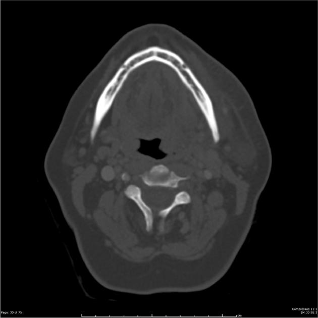
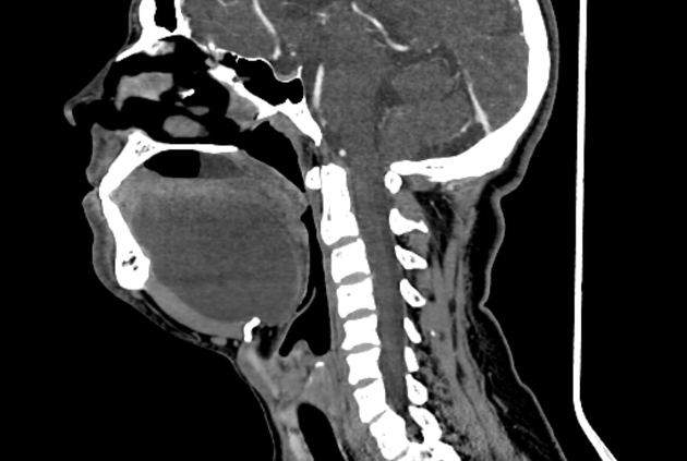
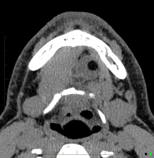
The floor of mouth is an oral cavity subsite and is a common location of oral cavity squamous cell carcinoma.
Posterior to the base of tongue inferior to the soft palate bounded laterally by the palatoglossal and palatopharyngeal arches and superior to the superior tip.
A prominent appearance of the pharyngeal fossa.
Ranulas present as a mass either in the floor of the mouth where they elevate the mucosa often with a blueish tinge or in the neck 7.
As the condition worsens the airway may be compromised with hardening of the spaces on both sides of the tongue.
It is stretching and displacing the genioglossus and mylohyoid muscles downwards.
This condition has a rapid onset over hours.
Communicates with the nasal cavity anteriorly.
It is composed of three parts.
The floor of the mouth is the part of the oral cavity that is located under the tongue.
It may be involved in a wide range of pathologic processes some of which are unique to the region.
Posterior to the nasal choanae extending from the vault of the pharynx superiorly to the soft palate inferiorly.
The lingual veins accompany the lingual artery between the hyoglossus and genioglossus and enters the internal jugular vein near the greater horn of the hyoid bone 3 the dorsal lingual vein which drains the dorsum and sides of the tongue joins the.