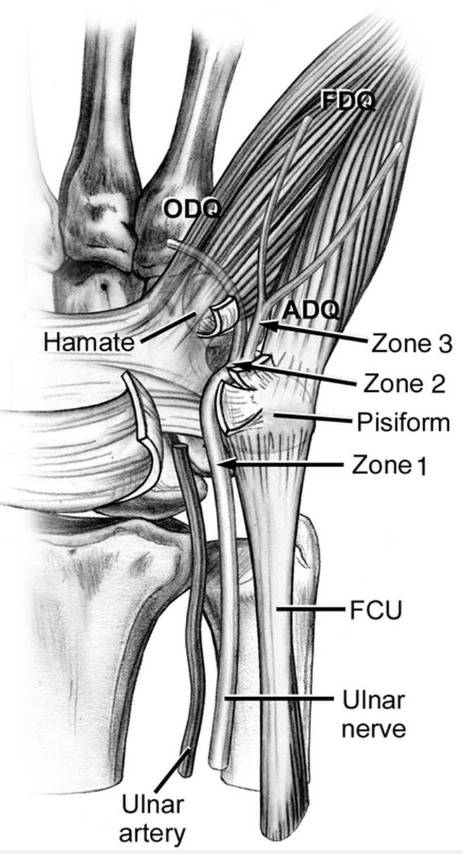
The ulnar tunnel is 4 4 5 cm in length and unlike the carpal tunnel the anatomic borders change from proximal to distal.
Floor of the distal ulnar tunnel.
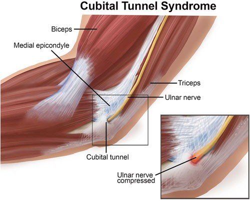
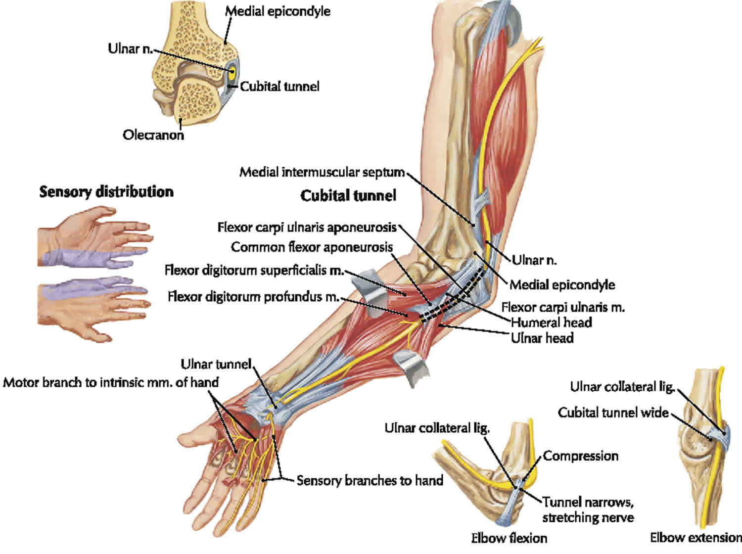
At elbow cubital tunnel humeral ulnar aponeurosis.
C8 and t1 c7 roots axons pass through brachial plexus.
Most distal to elbow.
4 the pisohamate ligament remains at the floor.
This anatomical space houses the ulnar nerve and ulnar artery as they pass from the distal forearm into the hand.
The tunnel begins at the proximal edge of the palmar carpal ligament and ends at the fibrous arch of the hypothenar muscles.
Compression of the ulnar nerve in the guyon canal is the fourth most common tunnel syndrome and a more common site of compression of the ulnar nerve is the cubital tunnel 2 3 4.
The ulnar nerve and artery run through guyon s canal.
Between pisiform hamate bones in hand branches.
The roof is formed by a fibrous arch of the hypothenar muscles abductor digiti minimi flexor digiti minimi brevis opponens digiti minimi and palmaris brevis listed from.
The distal ulnar tunnel was described by anatomist and urological surgeon felix guyon in 1861 based on his anatomical dissections investigating the unique and small protrusion of fatty tissue into the distal forearm noted when pressure was applied to the hypothenar eminence ollier phenomenon.
Nerve entry into wrist.
Under flexor carpi ulnaris guyon s canal.
Ulnar nerve the ecu at the distal ulna druj.
Lower trunk medial cord ulnar groove.
The distal ulnar tunnel was described by anatomist and urological surgeon felix guyon in 1861 based on his anatomical dissections investigating the unique and small protrusion of fatty tissue into.
Arises from lateral epicondyle of distal humerus passes through a fibro osseous tunnel as it leaves the f a 6th extensor compartment lies in a bony groove on dorsal surface of ulna inserts into base of 5th mc innervation.
Discussion the distal ulnar tunnel is a region of the wrist ap pro imately 4 cm in length in which the ulnar nerve is particularly vulnerable to external compression.
The tunnel is demarcated by the pisiform proximally and the hook of hamate distally.
The floor of this small tunnel is formed by the transverse carpal ligament its ulnar wall by the pisiform and its roof by the palmar carpal ligament which is an extension of the flexor retinaculum.
The ulnar tunnel is the space through which the ulnar neurovascular bundle passes at the wrist and within this confined space the nerve is susceptible to compression.