It is most commonly found in the submandibular salivary gland and duct followed by the parotid gland and its duct.
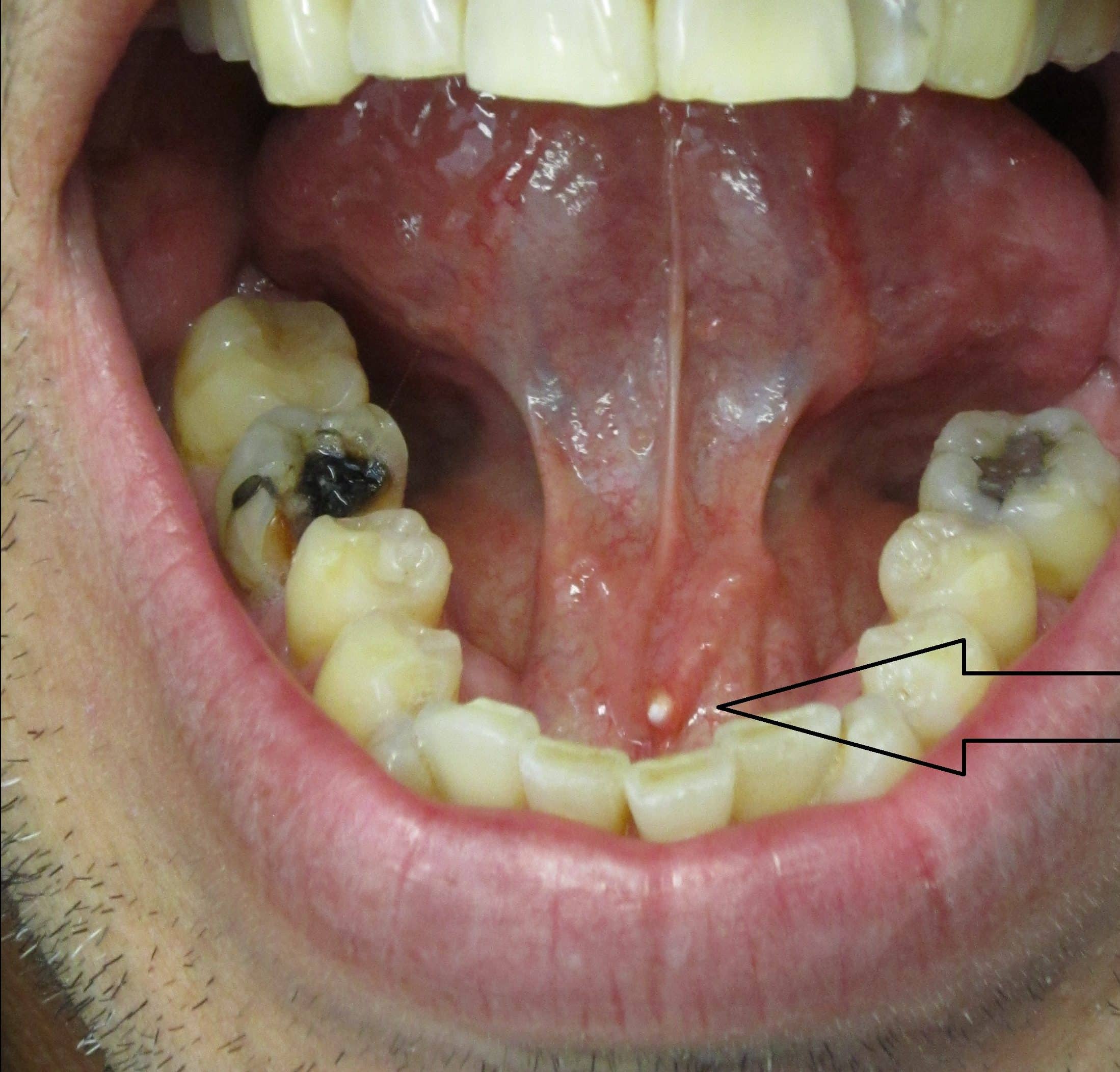
Floor of the mouth sialolith.
Causes range in severity including temporary irritation from food or beverages to sudden swelling that may tighten the throat and restrict breathing.
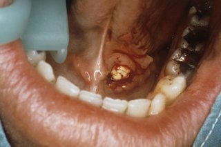
One of them had eroded through the floor of the buccal cavity forming an orocervical fistula and pus discharge.
It may be involved in a wide range of pathologic processes some of which are unique to the region.
A ranula is a mucus extravasation cyst involving a sublingual gland and is a type of mucocele found on the floor of the mouth.
The floor of the mouth is the part of the oral cavity that is located under the tongue.
The usual symptoms are pain and swelling of the.
The most common appearance is at the level of the submandibular gland this being also known as wharton s duct it is also possible however it only occurs rarely that the calculi appear at the level of the smaller salivary glands as well as in the sublingual or parotid gland.
The largest sialolith reported in the literature was 70 mm in the length of the wharton duct and was described as having a hen s egg size 2.
Less commonly the parotid gland or rarely the sublingual gland or a minor salivary gland may develop salivary stones.
The mucosal surface of the floor of the mouth is easily examined clinically as superficial abnormalities can be assessed visually without the aid of imaging.
Radiographic examination with a panoramic radiograph and occlusal radiograph revealed a radiopaque mass of size 3 1 cm extending anteroposteriorly and mediolaterally from the mandibular lateral incisor region to premolar region in the floor of the mouth suggestive of a sialolith figure 2.
Sialolithiasis is a medical condition in which calculi or stones form within the salivary glands.
Sialolithiasis is the abnormal formation of stones or salivary calculus in the salivary glands of a person.
Major and minor salivary glandspage contents1 major and minor salivary glands2 functions of.
Mouth swelling can happen in or around the mouth such as the roof of the mouth tongue and lips.
On the other hand asfar et al 7 reported three cases of giant submandibular intraglandular stones.
Read more below to learn about mouth swelling.
Ranulae present as a swelling of connective tissue consisting of collected mucin from a ruptured salivary gland caused by local trauma.
Sialolith located completely inside the submandibular gland that finally led to erosion of floor of the mouth.
Large sialoliths may perforate the floor of the mouth by ulcerating the duct or may result in orocutaneous fistula by causing a suppurative infection.
Sialolithiasis also termed salivary calculi or salivary stones is a condition where a calcified mass or sialolith forms within a salivary gland usually in the duct of the submandibular gland also termed wharton s duct.
Sublingual glands that are found beneath the tongue are the least affected.