Where medical information is easy to understand.
Floor of third ventricle mnemonic.
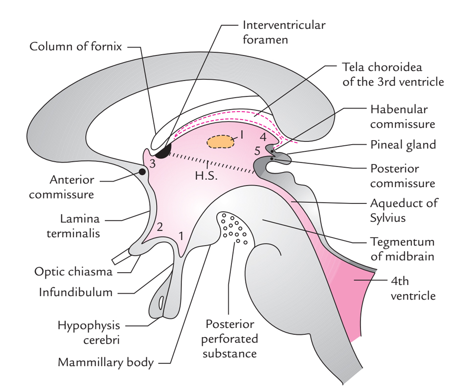
The third ventricle can be described as having six components.
This mnemonic stands for the above mentioned structures in the following order.
A handy mnemonic for remembering the structure of the floor of the third ventricle is tom pit tom the cat fell on the floor and went to a pit.
Posted by p12345 on 03 aug 2014.
The third ventricle is one of the four ventricles in the brain that communicate with one another.
The wall in the back is known as the posterior wall.
The third ventricle is surrounded by a number of structures of the diencephalon the diencephalon is a division of the forebrain that relays sensory information between brain regions and controls many autonomic functions.
As with the other ventricles of the brain it is filled with cerebrospinal fluid which helps to.
Its anterior wall is formed by the lamina terminalis columns of the fornix and the anterior commissure.
3445 people have seen this mnemonic.
Floor of 3rd ventricle form by.
Opin tuma posteg floor of third ventricle formed primarily by hypothalamic structures optic chiasma infundibular recess which extends into pituitary stalk tuber cinereum mammillary.
The walls on the side are known as the lateral walls.
In the sagittal plane it has a complex fishlike shape created by two anterior and two posterior recesses and a curving roof and floor most of the lateral walls of the ventricle are formed by the medial aspects of the two.
Floor of 3rd ventricle form by.
Ct and mr imaging anatomy of the third ventricle.
The third ventricle lies in the midline of the diencephalon.
In the axial and coronal planes it has a slitlike contour.
The third ventricle is one of the four csf filled cavities that together comprise the ventricular system.
Running through the third ventricle is the interthalamic adhesion which contains thalamic.
The third ventricle is one of the four connected ventricles of the ventricular system within the mammalian brain it is a slit like cavity formed in the diencephalon between the two thalami in the midline between the right and left lateral ventricles and is filled with cerebrospinal fluid csf.
3445 people have seen this mnemonic.
The third ventricle is a median cleft between the two thalami and is bounded laterally by them anteriorly and the hypothalamus and subthalamus posteriorly.
It links endocrine system nervous system and limbic system structures.
Floor of 3rd ventricle form by.
The third ventricle has a narrow roof a floor and four walls.
Third ventricle structure.